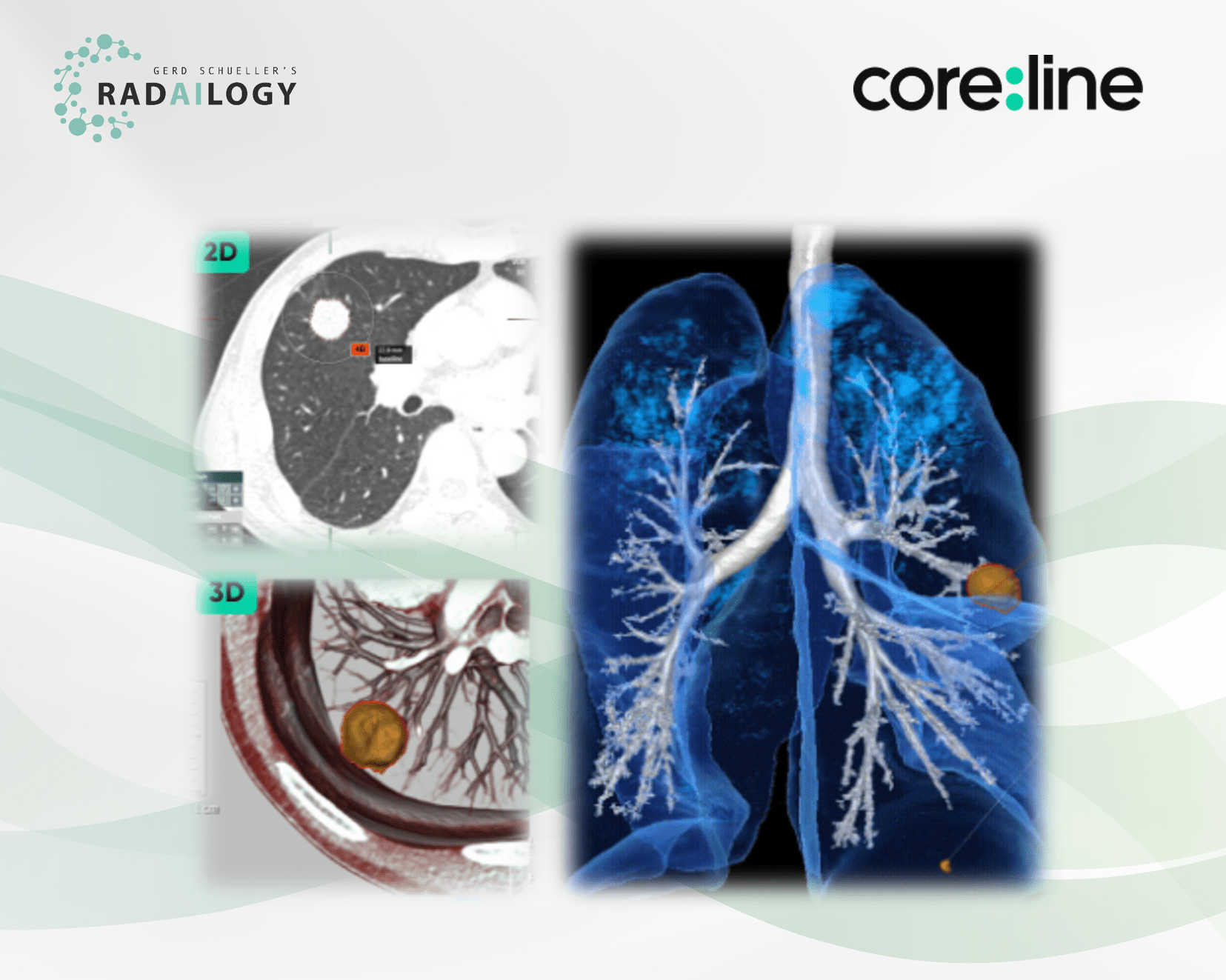
Non-enhanced low-dose chest CT of a 59-year-old male patient with lung cancer. A spiculated pulmonary nodule is visible at the base of the right upper lobe (upper left). aview LCS indicates the diameter, the volume as well as the morphology of the mass. In addition, the lesion is shown in clear 3D visualizations in relation to the vessels, the airways as well as the Interlobia (lower left and right).
Since lung nodules occur in more than two million people per year in Europe alone and the mortality rate from lung cancer worldwide is around two million annually, precise reporting microscopic nodules demands a variety of information, including the number of nodules, the size and status. In this setting, the strength of AI assistants is to reduce the radiologists´ workload and to allow highly accurate reports. In particular, providing high quality follow-up assessment shows the potential of AI assistants.
It is with great pleasure that we present Coreline´s aview LCS, an AI assistant for the detection of pulmonary nodules in CT studies.
Why aview LCS matters and how it works
The AI assistant detects and diagnoses lung nodules on chest CT studies. It provides 2D and 3D size and volume information by segmenting nodules, and automatically classifies solid, part-solid, non-solid. An important function is the automated comparison of nodules in the CT follow up. aview LCS reports according to the guidelines of the Lung CT Screening Reporting and Data System (Lung-RADS Version 1.1), as recommended by the American College of Radiology. The results are presented in tabular form and with 2D and 3D images. Each individual nodule is described with its exact location, diameter, volume and its morphology. The clear reports in words and images are a welcome support for the transfer of knowledge from radiologists to patients and clinicians. If aview LCS is used for general lung cancer screening, a workload reduction of between 77.4% and 86.7% can be expected according to the reference in our “The scientific evidence” section. This AI assistant can be used both for individual patients and in large oncology departments.
Who benefits
Patients, clinicians and radiologists by the reliable detection of pulmonary nodules with clear reports. In particular, the 2D and 3D visualization of the lungs and the nodules are a welcome help for interdisciplinary and patient communication.
Our own experience at Radailogy
Coreline reports the performance data as: sensitivity 97%, specificity 76%, accuracy 91%, ROC AUC .76, respectively. The AI assistant has high values for PPV, NPV and sensitivity of more than 92% each in our own patient population for lung nodules larger than 10mm. Lesions are precisely located and described using the parameters diameter, volume and morphology. Two CT studies are compared in the follow-up setting. The status of each nodule is indicated as baseline, unchanged, smaller or larger, respectively. New nodules are reported as such. This automated comparison in follow-up lung CTs reduces much of the workload. However, we could not validate the reduction of the workload by 86.7% and the time saving of 70% suggested by the vendor.
We also evaluated aview LCS together with aview COPD, which was developed to detect and quantify pulmonary emphysema in lung CT studies. Find out more in our AI assistant menu!
The scientific evidence
Lancaster HL, Zheng S, Aleshina OO, Yu D, Yu Chernina V, Heuvelmans MA, de Bock GH, Dorrius MD, Willem Gratama J, Morozov SP, Gombolevskiy VA, Silva M, Yi J, Oudkerk M. Outstanding negative prediction performance of solid pulmonary nodule volume AI for ultra-LDCT baseline lung cancer screening risk stratification. Lung Cancer. 2022 Jan 6;165:133-140.
Data to upload to Radailogy
Non-enhanced low-dose chest CT studies of any CT scanner; axial reformations; slice thickness and interval below 1.25mm each; sharp lung reconstruction kernel

