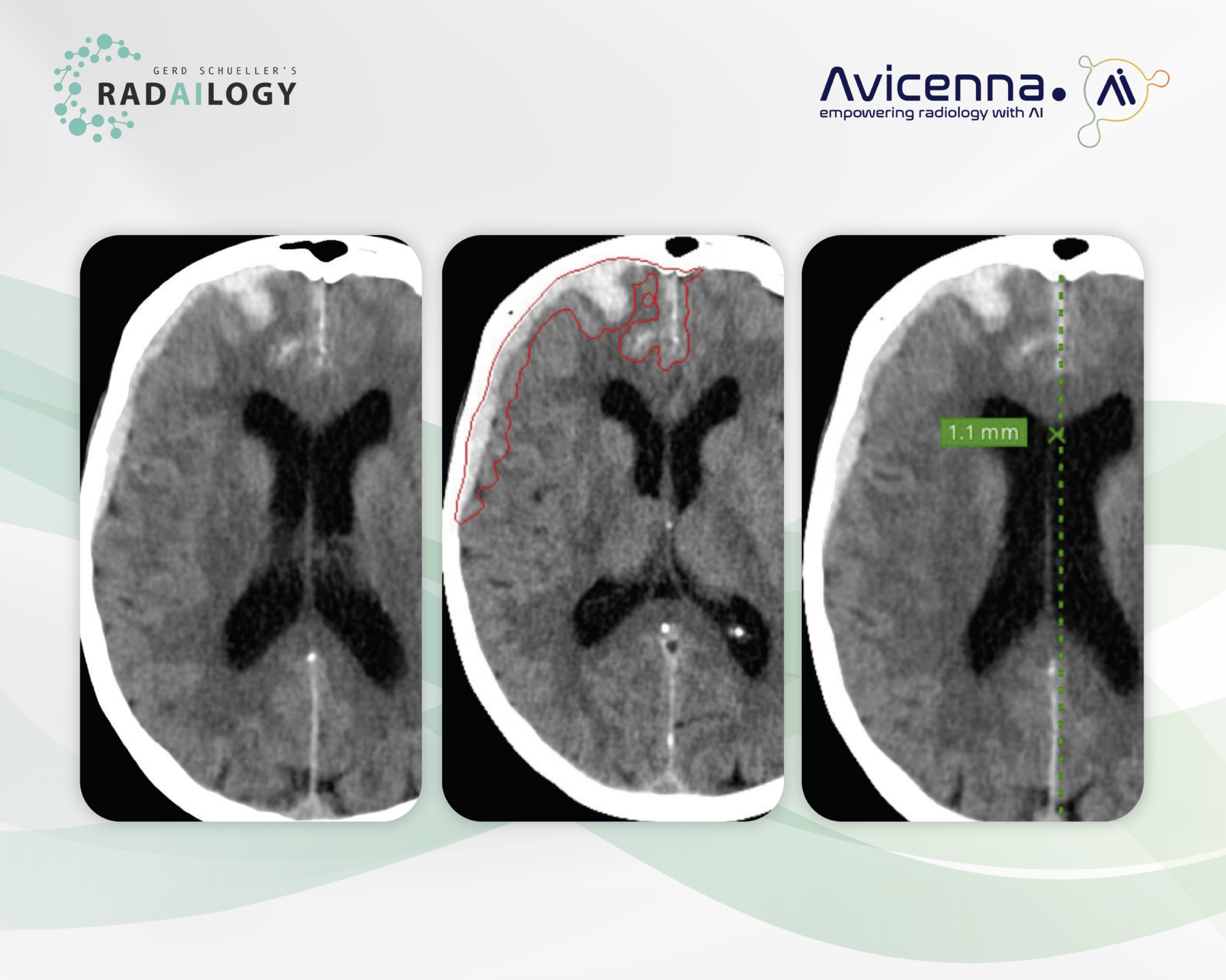
Non-enhanced cerebral CT of a 46-year-old patient after high-speed trauma. There is an acute subdural hemorrhage along the right frontal and parietal hemisphere (left). In particular, it is frontally surrounded by incipient edema and a slight contralateral midline shift can be seen. There is no intraventricular hemorrhage. There is no evidence of a parafalcine herniation.
CINA-ICH correctly and fully detects intracranial hemorrhage (middle). Bleeding volume is reported within the CT series and also tabulated (not shown). The slight contralateral midline shift is precisely measured (right).
Time is Health: Our motto is particularly understandable when it comes to head injuries and strokes. Every medical acute care unit knows what it means to accompany a large number of patients around the clock with the best diagnostic precision. Intracranial acute pathologies must be visualized quickly and safely.
It is with great pleasure that we present CINA-ICH, an AI assistant for intracranial hemorrhage detection in CT studies.
Why CINA-ICH matters and how it works
CINA-ICH was developed as a triage tool for emergency radiology. The AI assistant promptly reports suspected intracranial hemorrhages in CT scans and enables prioritization of these patients. The AI assistant provides radiologists and emergency physicians with an outstanding presentation of the findings in words and images.
Any medical professional can use CINA-ICH for each individual emergency patient by quickly and easily uploading cerebral CT studies to Radailogy. In medical institutions, this AI assistant can also do its work automatically in the background in order to fully benefit from its triage potential.
Who benefits
The triage of traumatic brain injury and hemorrhagic stroke is essential for everyone involved, i.e., patients, clinicians and radiologists.
Our own experience at Radailogy
We were able to study the performance of CINA-ICH in detail in extensive test series. On the one hand, we were convinced by the quick and clear presentation of findings and, on the other hand, by the low false-negative and false-positive rates in the detection of intracranial bleeding. This is consistent with the statistical data provided by the developer of sensitivity and specificity of more than 90% each, measured on the total cohorts.
The results are reported in detail. Bleeding is plotted in axial CT volumes. In addition, the data are processed in tabular form and include the parenchymal, sub- and epidural, subarachnoid and intraventricular bleeding volumes. Furthermore, midline shifts are measured.
The manufacturer describes the smallest bleeding with a volume of less than 3 ml and non-acute haemorrhage as a possible false-negative detection. At Radailogy, we have not observed any missed bleeding in our clinical testing.
We also evaluated CINA-ICH together with two other AI assistants, CINA-ASPECTS and CINA-LVO, which were developed to detect non-hemorrhagic strokes in CT studies. Find out more in our AI assistants!
The scientific evidence
McLouth J, Elstrott S, Chaibi Y, Quenet S, Chang PD, Chow DS, Soun JE. Validation of a Deep Learning Tool in the Detection of Intracranial Hemorrhage and Large Vessel Occlusion. Front Neurol. 2021 Apr 29;12:656112.
Rava RA, Seymour SE, LaQue ME, Peterson BA, Snyder KV, Mokin M, Waqas M, Hoi Y, Davies JM, Levy EI, Siddiqui AH, Ionita CN. Assessment of an Artificial Intelligence Algorithm for Detection of Intracranial Hemorrhage. World Neurosurg. 2021 Jun;150:e2 09 e217.
Data to upload to Radailogy
Non-enhanced cerebral CT studies of any CT scanner; axial reformations; minimal matrix size 256 x 256; maximal slice thickness 5 mm; tube current 100 kVp to 160 kVp (recommended 120 kVp to 140 kVp); soft tissue reconstruction kernel

