factsheet
aview COPD classifies and analyses COPD patterns on chest CT studies. An important feature is the tracking of disease in follow up CT studies. We consider aview COPD capable of shortening the radiologists´ workload while increasing the professional efficiency.
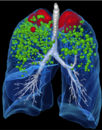
aview COPD demonstrates its full capacity to comprehensively assist in the diagnosis and visualization of the fundamental COPD pathologies in cross sectional imaging. aview COPD offers a variety of MPR images, diagrams and graphs to illustrate the pulmonary anatomy and disease as well as the distribution pattern of COPD. An interesting feature is the analysis and depiction of emphysema clusters using the D-slope value by applying a three-dimensional size-based emphysema clustering technique.
Non-enhanced low-dose chest CT studies of any CT scanner; axial reformations; slice thickness and interval 1.0 mm each; lung reconstruction kernel
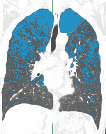
Non-enhanced low-dose chest CT of a patient with COPD. 2D image (left) and MPR visualization (right) of lung anatomy and emphysema clusters. Detailed charts and graphs indicated a multitude of morphological and pathological parameters and values (not shown).
Similar AI Assistants

aview CAC
aview CAC analyzes calcium in the coronary arteries and calculates the risk of a heart attack.
CT
Chest
Pixelshine
PixelShine generates significantly improved quality from these noisy images, for example in obese patients. Secondly, the lifespan of CT scanners is extended by reducing the load on the CT tubes.
CT
Multiple Organs

aview LCS
aview LCS detects and diagnoses lung nodules on chest CT studies. An important function is the automated comparison of nodules in the CT follow up.
CT
Chest

Rayscape Lung CT
Rayscape Lung CT identifies pulmonary nodules from 3 to 30 mm in diameter. An important function is the automated comparison of nodules in the follow-up from CT study to CT study.
CT
Chest


