factsheet
CINA-LVO reports suspected acute cerebral artery occlusions in CT angiographies and enables these patients to be prioritized. The assistant provides radiologists and emergency physicians with clear pictures.
Any medical professional can use CINA-LVO for each individual emergency patient by quickly and easily uploading CT angiographies of the skull base arteries to Radailogy. In medical institutions, this AI assistant can also do its work automatically in the background in order to fully benefit from its triage potential.
CINA-LVO (Large vessel occlusion) detects occlusions of the distal intracranial internal carotid artery and the M1 and M2 segments of the middle cerebral artery in CT angiographies of the skull base arteries. The suspected arteries are indicated in axial and coronal MIPs.
It is reasonable to use CINA-LVO together with CINA-ICH and CINA-ASPECTS, which were developed to detect hemorrhagic and non-hemorrhagic strokes in CT studies. Find out more in our AI assistants!
CT angiographies of the skull base arteries of any CT scanner; axial reformations; minimal matrix size 512 x 512; maximal slice thickness 1.25 mm; tube current 80 kVp to 140 kVp (recommended 120 kVp to 140 kVp); 100 to 400 mAs; artery reconstruction kernel
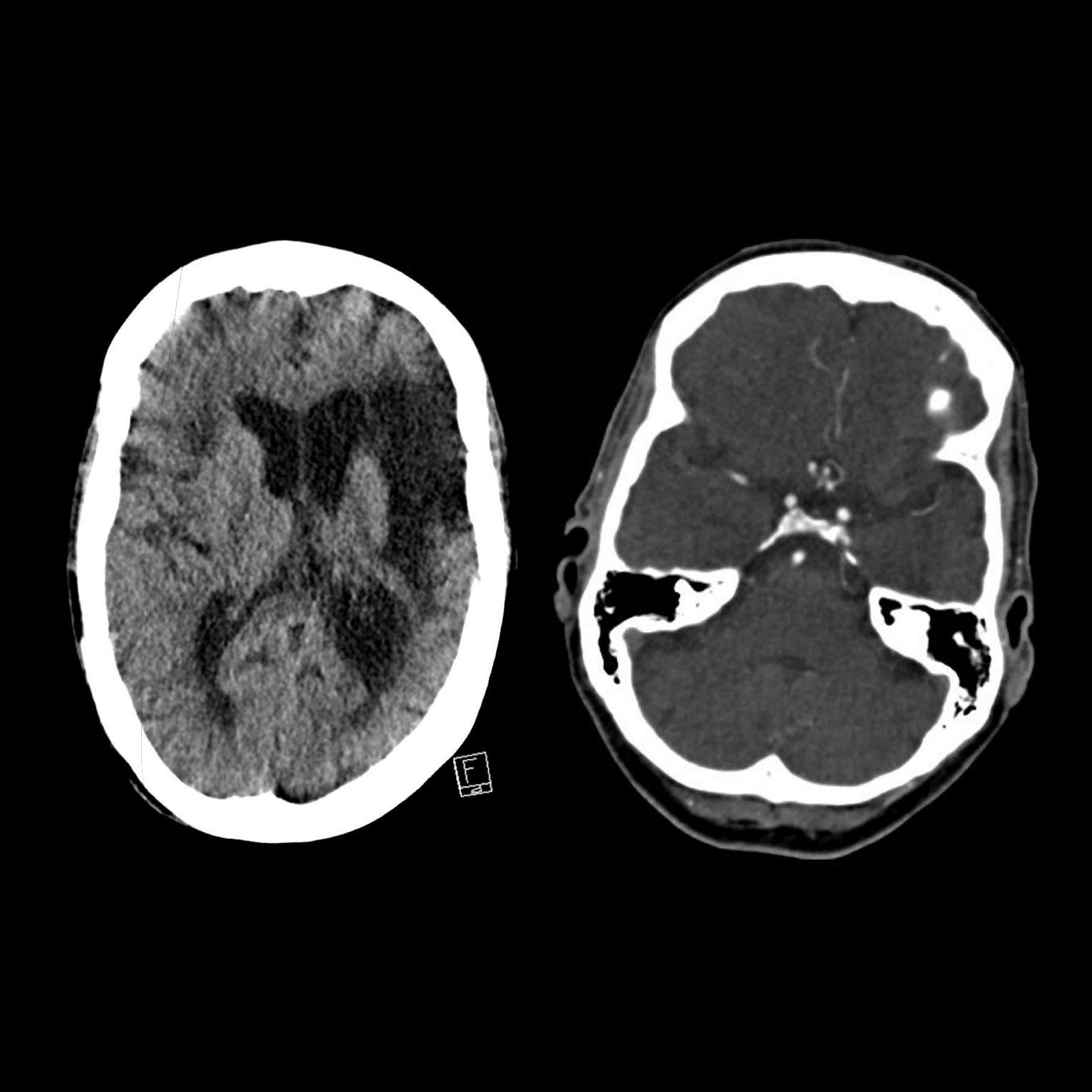
Cerebral CT of a 74-year-old patient with acute left sided weakness and known, pre-existing non-specific symptoms. Non-enhanced CT (left) shows moderate, older neurodegenerative changes in the supratentorial medulla and microangiopathic lesions in the deep gray matter on the right. There is discrete low attenuation in the lentiform nucleus and insular cortex and, questionably, in the posterior middle cerebral artery´s territory. CT angiography of the skull base arteries shows a contrast loss of the M2 segment of the right middle cerebral artery (right).
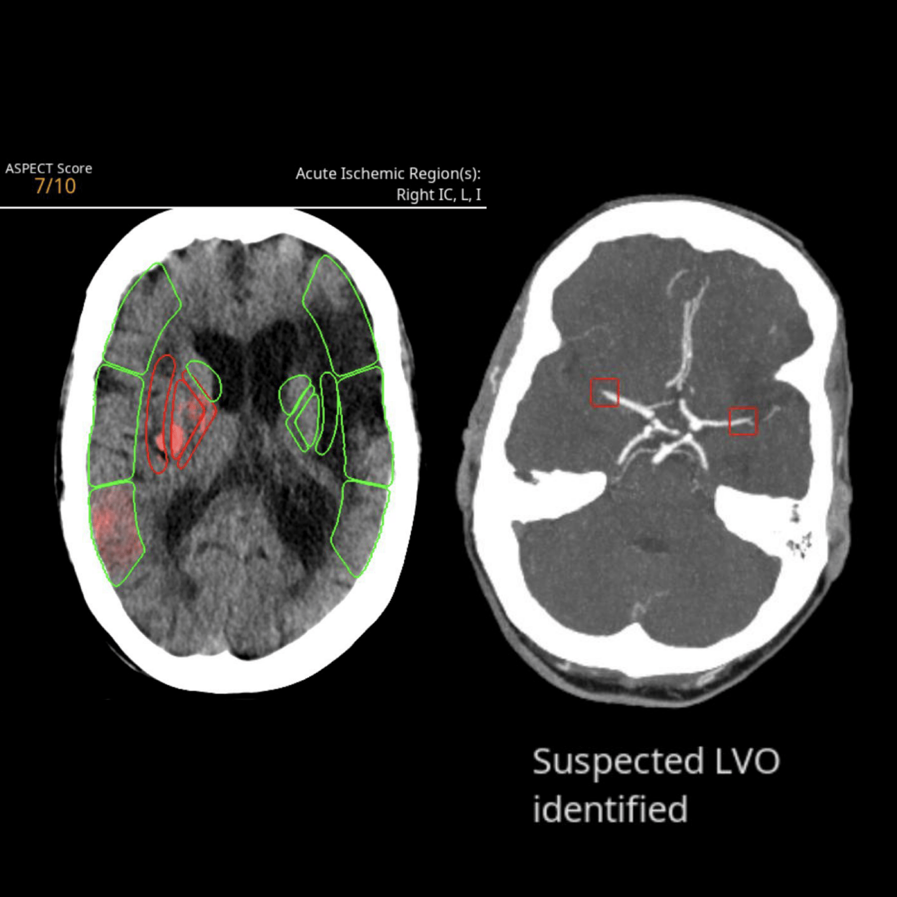
CINA-ASPECTS identifies the hypodensities and correctly assigns them to the right insula and the right basal ganglia (left, red). The low attenuation in the right posterior middle cerebral artery supply is detected and assigned to the M3 territory. However, they do not fall below the assitant's critical threshold for detecting acute ischemia. The ASPECTS is calculated as 8/10 (not shown). CINA-LVO correctly detects the artery occlusion (right, red box).
Similar AI Assistants

CINA-ASPECTS
CINA-ASPECTS detects acute intracranial ischemia in cerebral CT studies.
CT
Head and Neck

Brainscan CT
Brainscan CT reports acute and chronic intracranial processes in CT scans using tables and heat maps.
CT
Head and Neck

CINA-IPE
CINA-IPE detects acute pulmonary artery embolism in CT angiographies.
CT
Head and Neck
Pixelshine
PixelShine generates significantly improved quality from these noisy images, for example in obese patients. Secondly, the lifespan of CT scanners is extended by reducing the load on the CT tubes.
CT
Multiple Organs

CINA-ICH
CINA-ICH detects acute intracranial hemorrhage in cerebral CT studies.
CT
Head and Neck


