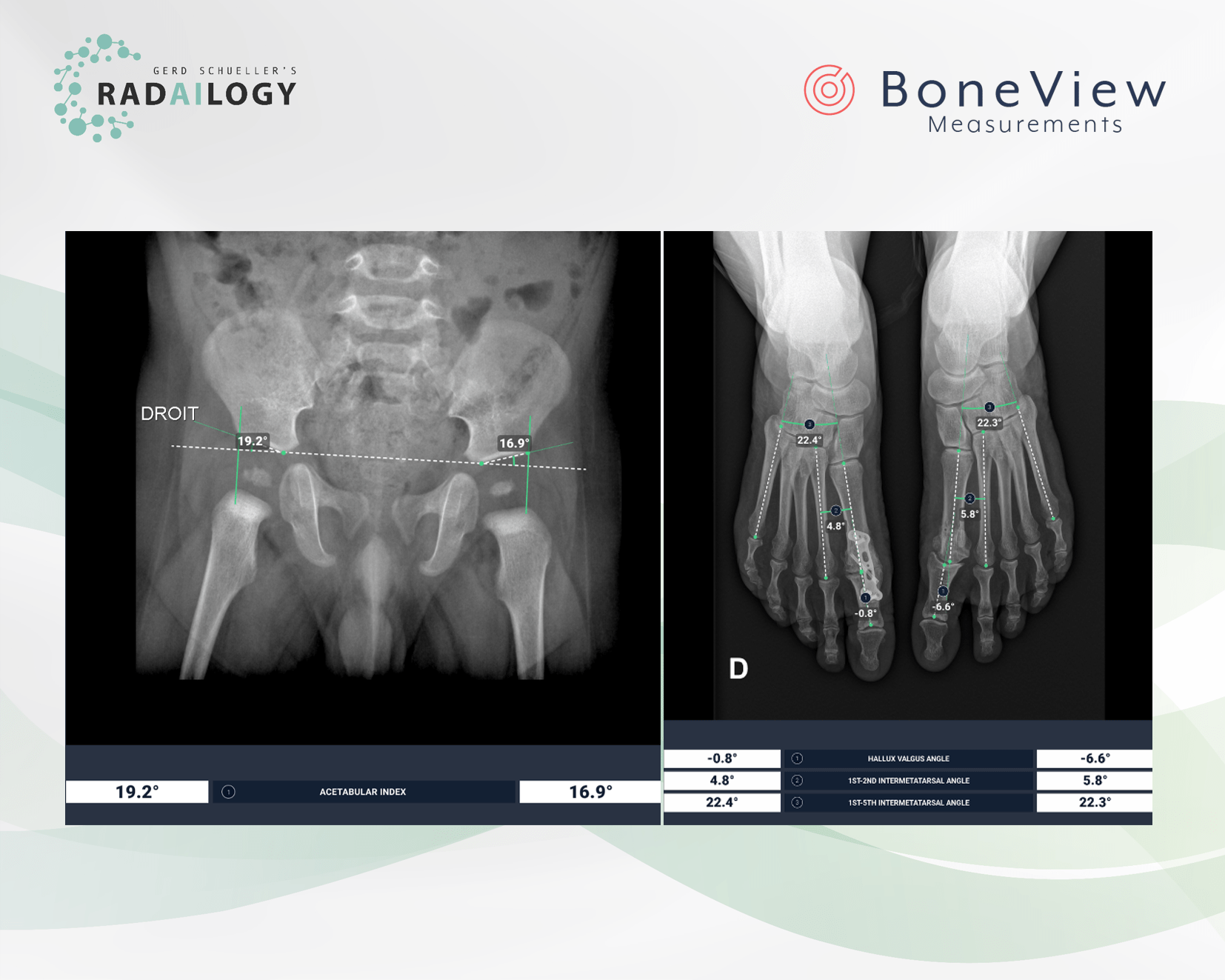
Radiography of the pelvis and both hip joints of a boy with the acetabular angles precisely measured on both sides by the AI assistant (left).
Frontal radiography of both forefeet of a 55-year-old patient. On the right (on the left in the picture) there are normal joint angles of the forefoot after osteomy of the first metatarsal bone. There is a hallux varus deviation on the left (on the right in the picture). The AI assistant measures the respective joint angles accurately.
We have the latest upgrade available for you! Gleamer´s BoneView Measurements of body axes come with interesting, new functions.
What is new?
Risser index
The Risser index is used to grade skeletal maturity for patients up to 18 years of age based on the degree of ossification and fusion of the iliac crest apophyses. It is primarily used as a marker of skeletal maturity, a surrogate for growth rate and potential, to plan scoliosis correction.
Acetabular angle for congenital hip measurement
The acetabular angle, or Sharp’s angle, is used to assess developmental hip dysplasia, particularly in patients with already ossifying epiphyses and therefore limited sonographic assessment.
Lumbar spine
The performance on lateral view measurements of the lumbar spine has significantly improved. Furthermore, lordosis is now calculated between L1 and S1, instead of between L1 and L5.
The scientific evidence
Hacquebord J, Leopold S. In Brief: The Risser Classification: A Classic Tool for the Clinician Treating Adolescent Idiopathic Scoliosis. Clin Orthop Relat Res. 2012;470(8):2335-2338.
Lee Y, Chung C, Koo K, Lee K, Kwon D, Park M. Measuring Acetabular Dysplasia in Plain Radiographs. Arch Orthop Trauma Surg. 2011;131(9):1219-1226.
Data to upload to Radailogy
Digital radiography of the skeleton

