News
Discover the World of AI
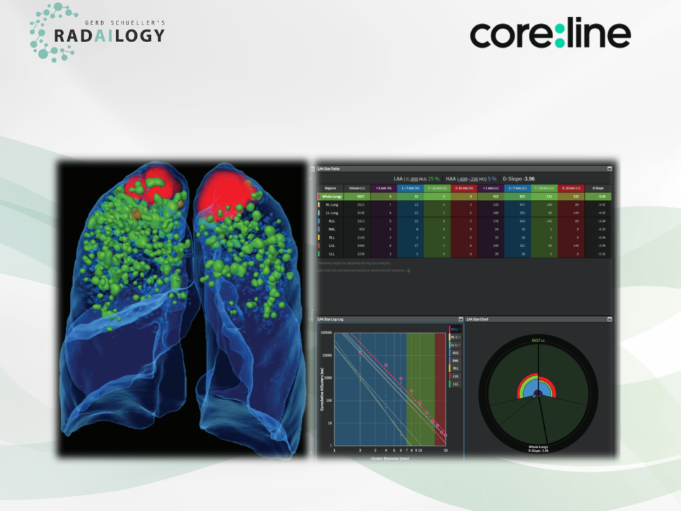
CT and COPD: AI with previously unknown potential
Non-enhanced low-dose chest CT of a patient with COPD. MPR visualization of lung anatomy and emphysema clusters (left) and detailed charts and graphs (right). The absolute volumina and the relative low attenuation volumina (25%) were given for both l ...
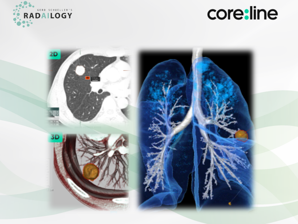
Lung CT: Reduce your workload and increase your diagnostic accuracy!
Non-enhanced low-dose chest CT of a 59-year-old male patient with lung cancer. A spiculated pulmonary nodule is visible at the base of the right upper lobe (upper left). aview LCS indicates the diameter, the volume as well as the morphology of the ma ...
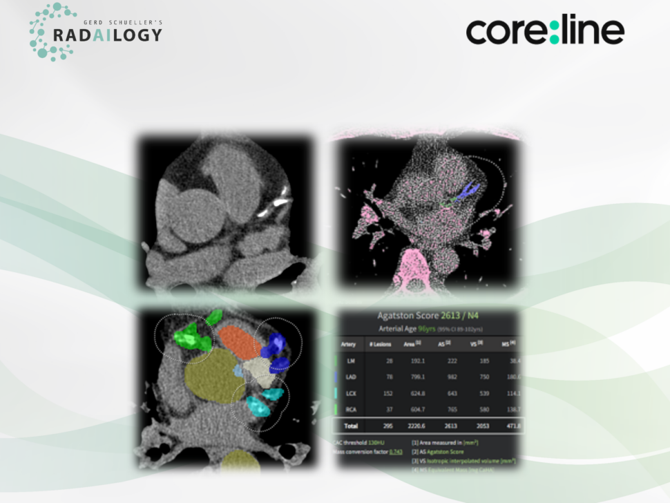
Do you know your risk of heart attack? Done quickly with aview CAC!
Non-enhanced chest CT of a 65-year-old patient; 120 Kvp; axial reformation; soft tissue reconstruction kernel. aview CAC segments all cardiac structures and detects coronary artery calcium in all coronary arteries, calculates an Agatston score of 261 ...
For everyone who can still be amazed: meet us at the ECR 2025!
Radailogy and ERS at the ECR! Meet us in Hall X1 Booth A01 from February 26 to March 1, 2025!Our world of Artificial Intelligence and Teleradiology up closeOne of our great strengths is that we listen carefully to our partners and understand exactly ...
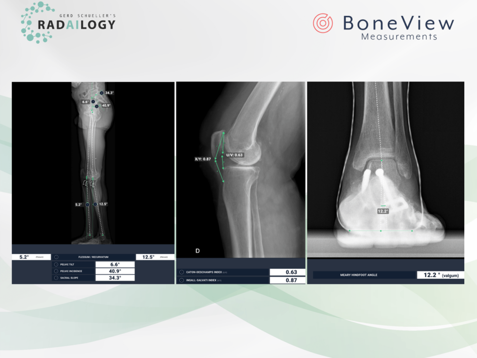
New features for automated measurements of body axes on X-ray images: Part 2
Lateral radiographs of both legs (left) and the right knee joint (middle), radiography of the left ankle and forefoot a.p. (right). BoneView Measurements calculates the iliosacral body axes, the pelvic tilt and the femorotibial joint angles to determ ...
New features for automated measurements of body axes on X-ray images: Part 1
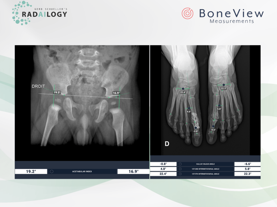
Radiography of the pelvis and both hip joints of a boy with the acetabular angles precisely measured on both sides by the AI assistant (left).Frontal radiography of both forefeet of a 55-year-old patient. On the right (on the left in the picture) t ...
Acute and chronic pathologies at a glance: precise CT diagnostics of the brain
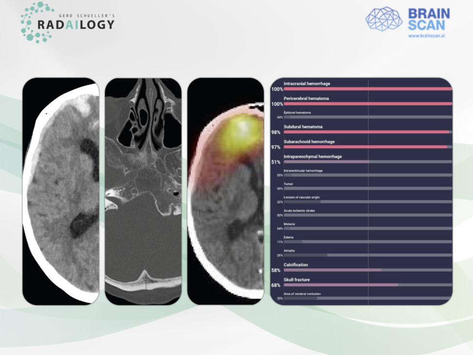
Non-enhanced cerebral CT of a 61-year-old patient after a motor vehicle accident. There is acute intraparenchymal, subdural, and subarachnoid hemorrhage along the right hemisphere (left) and a nondisplaced occipital skull fracture (middle left).Brain ...
MRI of the prostate: Convincing detection and analysis of prostate cancer
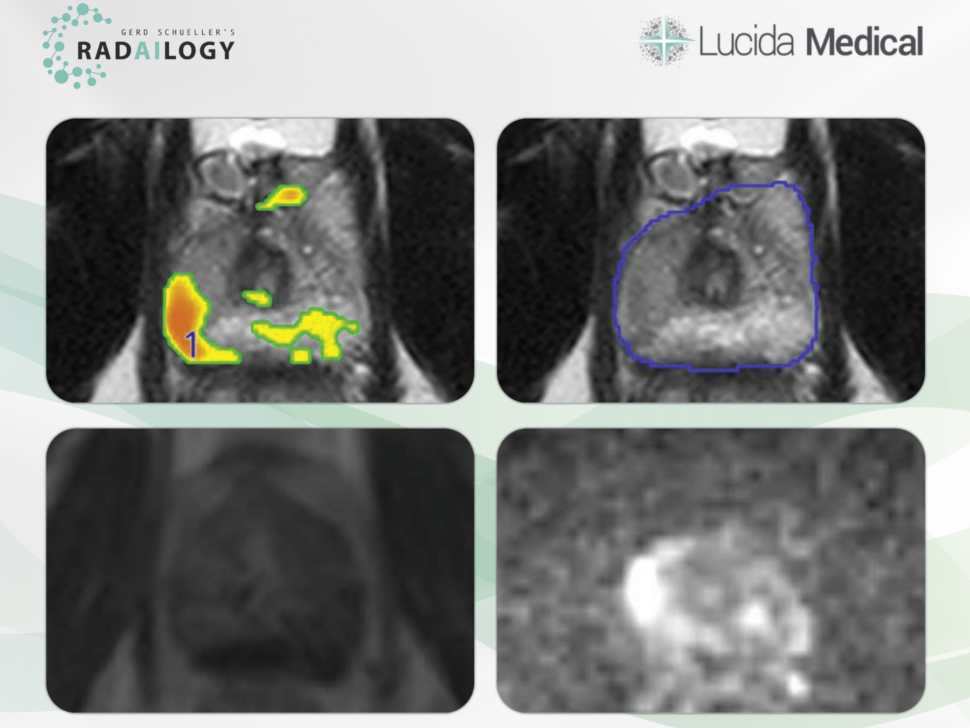
Axial T2 (upper left and right), contrast enhanced T1 (DCE, lower left) and DWI (lower right) MRI of the prostate of a 52-year-old patient with a Gleason Score of 4. T2 hypointensity in the middle peripheral zone on the right with corresponding hyper ...

