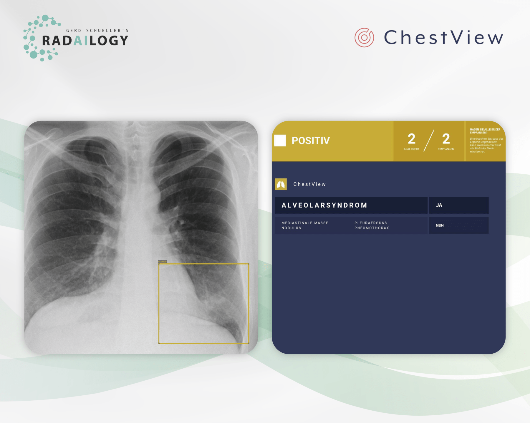
Chest radiograph of a 39-year-old patient (left) with symptoms of a lower respiratory tract infection. Small lung opacities are visible in projection to the left lower lung lobe. ChestView correctly identifies the pathology (yellow box). The result is also listed in a table (right). In addition to the fully automatic notification of the acute pathology, other threatening pathologies of the thorax are correctly evaluated as negative.
Chest radiography is used worldwide as one of the most common, if not the most common, radiological examinations in both acute care and elective medicine. Correct and reproducible reports and the communication of results from radiologists to patients and clinicians are of the utmost importance for everyone involved.
It is with great pleasure that we present ChestView, an AI assistant for chest radiography.
Why ChestView matters and how it works
ChestView is an expert-level AI assistant that has been extensively tested thanks to the joint work of our multidisciplinary team of developers and radiologist.
With ChestView essential pathologies of the thorax are diagnosed. The AI assistant was developed on the one hand to support triage in acute medicine and on the other hand to improve radiological work in terms of time savings and increased accuracy.
Who benefits
Patients, clinicians and radiologists by identifying the most important chest diseases with pictures and tables.
Our own experience at Radailogy
ChestView supports the detection of major thoracic pathologies. Although pneumonia is referred to as alveolar syndrome, the differential diagnosis has worked with a high degree of accuracy in our detailed tests. The presentation of the results by means of boxes and tabular description is helpful for radiological knowledge transfer to clinicians and patients.
The scientific evidence
Bennani S, Regnard NE, Lassalle L, Nguyen T, Malandrin C, Koulakian H, Khafagy P, Chassagnon G, Revel MP. Evaluation of radiologists’ performance compared to a deep learning algorithm for the detection of thoracic abnormalities on chest X-ray. In press
Data to upload to Radailogy
Digital radiographs of the chest for patients aged 15 and older

